

The Skin e-chart: Full illustrated
The Skin e-chart: Full illustrated by HC-HealthComm
English | May 25, 2016 | ISBN: N/A | ASIN: B01G7LS3SG | EPUB | 0.64 Mb
English | May 25, 2016 | ISBN: N/A | ASIN: B01G7LS3SG | EPUB | 0.64 Mb
The Skin e-chart, Full illustrated
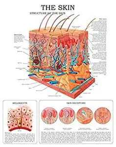
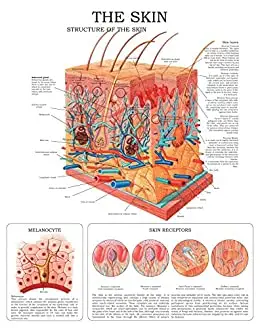
Structure of the skin
Skin layers
The skin is made up of three layers. the most superficial one is called epidermis; the middle layer is called dermis, and hypodermis the one underneath.
Melanocyte
This picture shows the cytoplasmic process of a melanocyte, which contains the melanin grains transferred to the interior of the cytoplasm of the epidermal cells in order to provide complexion to the skin. Melanin is a dark brown pigment, also responsible for the color of hair and eyes. An excessive exposure to UV rays can make the genetic material mutate and turn a normal cell into a cancerous one.
Skin receptors
Epidermis the thickness of this region varies according to the area under study. the most complex part is the one in the sole of the foot and in the palm of the hand, where thickness can amount to 1.5 mm. its epithelium is pluristratified (5 layers or cellular strata), and is made up of keratin laminae.
Keratinocytes are replaced by means of the division (mitosis) of basilar cells (regeneration). this skin layer also contains cells with pigments called melanocytes that are in charge of skin complexion; langerhansґ cells, which provide defensive functions and nerve cells, responsible for hormonal functions (merkelґs cells). it does not have blood vessels.
Dermis. It has variable thickness, which amounts to 3 mm. in the sole of the foot. It is a conjunctive tissue upon which the epidermis lies, consisting mainly in (collagen) fibers, connective tissue cells (fibroblasts), immunologically active phagocytes (macrophages), and mast cells that mediate allergic and inflammatory reactions. this dermal layer contains blood and lymphatic vessels, as well as sensitive receptors, hair, and sebaceous and sudoriferous glands.
The sudoriferous glands produce an acidic secretion acting as a protective layer that does not allow bacterial growth on the skin.
Hypodermis. It is made up of lax conjunctive tissue, slightly joining the dermis with the underlying organs. it is also made up of a variable layer of adipose tissue with an isolating function that allows the skin to modify and protect itself against the loss of heat and superficial traumas.
Feel Free to contact me for book requests, informations or feedbacks.
Without You And Your Support We Can’t Continue
Thanks For Buying Premium From My Links For Support
Without You And Your Support We Can’t Continue
Thanks For Buying Premium From My Links For Support