

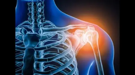
Radiology X-Ray Of Shoulder Joint

Radiology X-Ray Of Shoulder Joint
Published 11/2024
MP4 | Video: h264, 1920x1080 | Audio: AAC, 44.1 KHz
Language: English | Size: 295.36 MB | Duration: 2h 0m
Published 11/2024
MP4 | Video: h264, 1920x1080 | Audio: AAC, 44.1 KHz
Language: English | Size: 295.36 MB | Duration: 2h 0m
Master Shoulder Joint X-ray Interpretation: From Anatomy to Advanced Pathology"
What you'll learn
Master Core X-Ray Views: Understand and interpret essential shoulder X-ray views, including AP, true AP, Stryker notch, lateral Y, Neers, axial, and Garth views
Recognize and Differentiate Anatomical Landmarks: Identify key anatomical structures in each view, with an emphasis on detailed shoulder anatomy
Identify Pathologies and Traumatic Injuries: Detect common and complex shoulder injuries, such as fractures (e.g., greater tuberosity, humeral neck) dislocation
Interpret Degenerative and Systemic Conditions: Analyze radiographic signs of shoulder pathologies like osteomyelitis, rheumatoid arthritis neuropathicarthritis
Apply Checklists to Ensure Comprehensive Analysis: Use structured checklists for AP and apical oblique views to systematically review shoulder X-rays.
Diagnose Pediatric and Developmental Abnormalities: Identify conditions like Salter-Harris fractures, Sprengel deformity, cleidocranial dysostosis,
Understand Common Pitfalls and Misinterpretations: Recognize pseudo-lesions, pitfalls in dislocation interpretation, and potential errors
Learn to identify various bone lesions, including osteochondromas, chondroblastomas, ABCs, SBCs, and osteosarcomas.
Evaluate shoulder injuries using specialized views, such as ACJ, SCJ, and injury views, to gain a complete assessment of trauma cases.
Apply learning in quizzes and case studies to reinforce skills and prepare for clinical practice.
Requirements
Basic Anatomy Knowledge: A foundational understanding of human anatomy, especially musculoskeletal anatomy, will be helpful for following the detailed anatomical discussions.
Familiarity with Medical Terminology: Since the course includes specific radiological and anatomical terms, a general familiarity with medical terminology is recommended.
Interest or Background in Radiology or Medicine: This course is ideal for radiology students, medical students, or healthcare professionals interested in diagnostic imaging and shoulder radiography.
No Prior X-ray or Radiography Experience Needed: While some background in radiology is beneficial, the course is structured to guide you from basic to advanced topics, making it suitable for beginners as well as more experienced individuals
Description
Welcome to "Comprehensive Shoulder Joint X-ray Radiography," your go-to course for mastering the intricacies of shoulder X-ray interpretation. This course is designed to equip you with the knowledge and skills needed to confidently read and analyze shoulder X-rays, from basic views to complex pathologies.You’ll begin with an introduction to key shoulder X-ray views, such as AP, true AP, Stryker notch, lateral Y, Garth (apical oblique), and Neer’s view, as well as specialized views for the clavicle and AC joint. Through each section, you'll learn optimal positioning techniques, discover checklists for accurate interpretation, and gain the ability to identify critical structures and ossification centers.The course dives deep into pathology, helping you recognize and interpret various shoulder injuries and conditions, such as:Fractures (e.g., humeral head, glenoid rim, clavicle, scapula)Dislocations (anterior, posterior, inferior, ACJ, SCJ)Soft tissue injuries, including rotator cuff tears and calcific tendonitisArthritis types, osteomyelitis, AVN of the humeral head, and moreYou’ll also explore developmental abnormalities and tumor-related pathologies, learning to distinguish these from normal anatomy to avoid diagnostic pitfalls.Each section includes practical exercises, case studies, and quizzes, allowing you to apply your knowledge in real-world scenarios. By the end of the course, you’ll have a thorough understanding of shoulder radiography, enabling you to interpret shoulder X-rays with accuracy and confidence in a clinical setting.Who This Course Is For:Radiology residents, medical students, radiologists, and radiographersOrthopedic and sports medicine professionalsEmergency medicine practitionersMedical imaging technologists and allied health workersJoin this course today and take a step closer to becoming proficient in shoulder joint X-ray radiography!
Overview
Section 1: Introduction
Lecture 1 Introduction
Section 2: Positions in shoulder joint
Lecture 2 AP view
Lecture 3 True AP
Lecture 4 Stryker notch view
Lecture 5 Lateral Y view
Lecture 6 Neers view
Lecture 7 Axial view
Lecture 8 Apical oblique view
Lecture 9 Clavicle view
Lecture 10 ACJ view
Lecture 11 SCJ view
Lecture 12 Views if you suspect injury
Section 3: Anatomy of shoulder joint
Lecture 13 Anatomy in AP view
Lecture 14 Apical oblique anatomy
Lecture 15 Neers view anatomy
Lecture 16 Anatomy of axial view
Lecture 17 ACJ anatomy
Lecture 18 Checklist in AP view
Lecture 19 Checklist in apical oblique view
Lecture 20 Ossification centers
Lecture 21 Pitfall in shoulder
Lecture 22 Anatomy of conoid tubercle
Lecture 23 CC joint
Section 4: Variants
Lecture 24 Humeral Head pseudo lesion
Lecture 25 Os acromial
Lecture 26 Shape of acromion
Section 5: Trauma
Lecture 27 Traumatic shoulder
Lecture 28 Greater tuberosity fracture
Lecture 29 Humerus head neer fracture
Lecture 30 Humerus Neck fracture
Lecture 31 Child with salter harris type 1
Lecture 32 Fracture with bad prognosis
Lecture 33 Glenoid rim fracture
Lecture 34 Shaft fracture
Lecture 35 Clavicle fracture
Lecture 36 Scapula fracture
Lecture 37 Anterior shoulder dislocation
Lecture 38 Pitfall in dislocation
Lecture 39 Fracture with anterior dislocation
Lecture 40 Posterior dislocation
Lecture 41 Dislocation pitfall
Lecture 42 Inferior dislocation
Lecture 43 ACJ dislocation
Lecture 44 SCJ injury
Section 6: Non trauma
Lecture 45 Osteoarthritis
Lecture 46 Rheumatoid arthritis
Lecture 47 Neuropathic arthritis
Lecture 48 Rotator cuff injury
Lecture 49 Calcific tendonitis -bursitis
Lecture 50 Osteochondromatosis
Lecture 51 CPPD
Lecture 52 AVN humerus Head
Lecture 53 Distal clavicle osteolysis
Lecture 54 Osteomyelitis in neonate
Lecture 55 Chondroblatoma
Lecture 56 ABS and SBC
Lecture 57 Osteosarcoma
Lecture 58 Sprengle deformity
Lecture 59 Cleidocranial dysostosis
Lecture 60 Answers of quiz
Radiologists and Radiographers who want a refresher on shoulder imaging techniques and common pathologies.,Radiology Residents and Medical Students looking to strengthen their understanding of shoulder radiography and gain practical skills in X-ray interpretation.,Orthopedic Residents and Surgeons interested in honing their skills in identifying fractures, dislocations, and other shoulder injuries on X-rays.,Emergency Medicine and Sports Medicine Practitioners who frequently handle shoulder injuries and need a deeper knowledge of X-ray interpretation for immediate clinical decision-making,Healthcare Professionals and Allied Health Workers involved in patient imaging who would like to gain more insight into shoulder X-ray anatomy, views, and common findings.,Medical Imaging Students and Technologists aiming to expand their knowledge of shoulder radiographic techniques, anatomy, and pathology as part of their professional training.