


Radiographic (X-Ray) Positioning Made Easy: Lower Extremity.
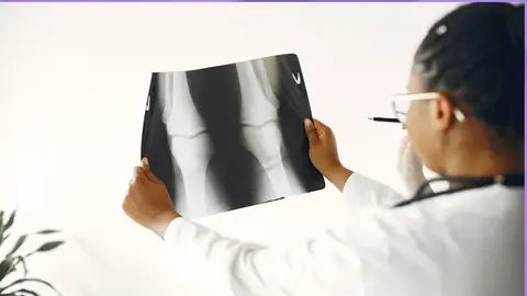
Radiographic (X-Ray) Positioning Made Easy: Lower Extremity.
Published 6/2023
MP4 | Video: h264, 1280x720 | Audio: AAC, 44.1 KHz
Language: English | Size: 1.31 GB | Duration: 1h 15m
Published 6/2023
MP4 | Video: h264, 1280x720 | Audio: AAC, 44.1 KHz
Language: English | Size: 1.31 GB | Duration: 1h 15m
Learn How to Position a Patient for X-ray of different parts of LOWER EXTREMITY: FOOT, ANKLE, CALCANEUM, KNEE, FEMUR…
What you'll learn
Medical Imaging: How To Position Patients for Lower Limb X-rays.
Radiology: Foot, Toes, Ankle, Calcaneum, Tibia & Fibula, Knee, Femur X-ray techniques.
You will become confident with positioning of patients for X-ray examinations.
Complete training and be confident with Upper Limb X-rays, Anatomy and Radiographs.
Requirements
No Previous Experience Needed.
Description
When I was a student radiographer at Canterbury Christ Church University here in England, I struggled with radiographic positioning. Most times in clinical placements radiographers were very busy with heavy work loads and had little time to spend explaining how to position a patient for an X-ray. Most times they were very quick and before I could figure out what they were doing the X-ray had already been done.Hence, I'm teaching this course from the student perspective, by-passing all the jargons to make sure I teach you exactly what you need to know to thrive as an excellent and confident radiographer. Radiographic Positioning Made Easy is not filled with high technical, text-book terminologies, but simple and direct skills of what I have learnt from my British National Health Service (NHS) experience as a qualified practicing radiographer, with over 35,000 patients seen in the last six years.I am sure that there are many student radiographers out there who are going through exactly what I went through some years back as an undergraduate student. If you are that kind of person then you are in the right place. At the end of this course you will be very confident and bold about your positioning.I applied a systematic approach to this course and logically divided the course into different body areas:Each section has different lectures covering specific body parts. Upper Extremity is made up of the following lectures viz:Lecture 1- IntroductionLecture 2 - Welcome messageLecture 3 - How to position patients for a FOOT X-rayLecture 4 - How to position patients for TOES X-rayLecture 5 - How to position patients for BIG TOE ? Hallux ValgusLecture 6 - How to position patients for an ANKLE X-rayLecture 7 - How to position patients for CALCANEUM X-rayLecture 8 - How to position patients for TIBIA & FIBULA X-rayLecture 8 - How to position patients for a KNEE X-rayLecture 9 - How to position patients for FEMUR X-rayLecture 10 - How to position patients for FEMUR KNEE DOWN X-ray.Lecture 11 - How to position patients for FEMUR KNEE UP X-ray Lecture 12 - ConclusionIn this way it's easy for you to go directly and get the specific information you need at any given time without wasting your precious time looking through endless search engine results.Furthermore, it's important to note that Radiography is the backbone of modern healthcare and positioning of the patient is the heart of any radiographic examination. It is the core of what we do as radiographers. If you get positioning wrong, you might end up with a sub-optimal image or even a non-diagnostic image which is not really what you want. Radiographic positioning is vital important whether you are using Computed Radiography (CR) or Digital Radiography (DR) systems.At the end of each lecture there is a short assignment to make sure you are abreast with the key skills. I am sure that at the end of this course you will be bold and confident to carry out different radiographic examination.Finally, I am always happy to assist you in any area that you need more help. Please feel free to leave a comment here or connect with me on social media through my Udemy personal profile.Happy Learning!!!
Overview
Lecture 1 Introduction
Lecture 2 Welcome Lecture
Section 1: X-ray Positioning: FOOT, TOES & TOE Lateral (Hallux Valgus)
Lecture 3 FOOT
Lecture 4 TOES
Lecture 5 TOE Lateral (HALLUX VALGUS)
Section 2: X-ray positioning: ANKLE, CALCANEUM, TIBIA & FIBULA.
Lecture 6 ANKLE
Lecture 7 CALCANEUM
Lecture 8 Tibia and Fibula
Section 3: X-ray positioning: KNEE, FEMUR KNEE DOWN, FEMUR KNEE UP.
Lecture 9 KNEE
Lecture 10 FEMUR KNEE UP
Lecture 11 KNEE DOWN
Section 4: CONCLUSION
Lecture 12 CONCLUSION
Student Radiographers, Newly Qualified Radiographers, Radiologic Technologist Students, A-level students, High School students.