


Head And Neck Neuroanatomy
Head And Neck Neuroanatomy
MP4 | Video: h264, 1280x720 | Audio: AAC, 44.1 KHz
Language: English (US) | Size: 489.76 MB | Duration: 0h 32m
MP4 | Video: h264, 1280x720 | Audio: AAC, 44.1 KHz
Language: English (US) | Size: 489.76 MB | Duration: 0h 32m
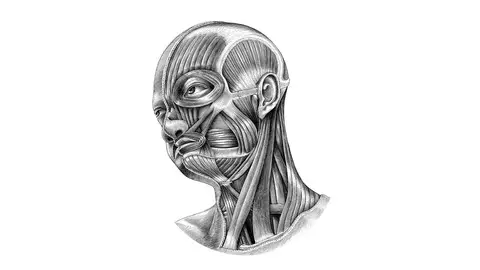
study of major sub branches of Head and Neck Neuroanatomy which includes Major Cranial Nerves etc.
What you'll learn
trigeminal nerve
facial nerve
anterior cranial fossa
middle cranial fossa
ventricles of brain
Requirements
medical student
dentistry student
Description
This course includes knowledge about the Trigeminal Nerve, Facial Nerve, ventricles of the brain, anterior cranial Fossa, and middle cranial fossa. the student will get to know about the significance and clinical relevance of these structures. Their importance and Core knowledge about it. Students will Surely Enjoy this Course for their Head and Neck Anatomy.The head and neck is covered in skin and its appendages, termed the integumentary system. These include hair, sweat glands, sebaceous glands, and sensory nerves. The skin is made up of three microscopic layers: epidermis, dermis, and hypodermis. The epidermis is composed of stratified squamous epithelium and is divided into the following five sublayers or strata, listed in order from outer to inner:Stratum corneum,Stratum lucidum,Stratum granulosum,Stratum spinosum,Stratum germinativum also called stratum basale. The deepest layer is the miotic layer, stratum basale producing daughter cells by mitosis.The brachiocephalic artery or trunk is the first and largest artery that branches to form the right common carotid artery and the right subclavian artery. This artery provides blood to the right upper chest, right arm, neck, and head, through a branch called right vertebral artery. The right and left vertebral artery feed into the basilar artery and upward to the Posterior cerebral artery, which provides most of the brain with oxygenated blood. The posterior cerebral artery and the posterior communicating artery are within the circle of Willis.The left common carotid artery divides to form the: internal carotid artery (ICA) and an external carotid artery (ECA). The ICA supplies the brain. The ECA supplies the neck and face.The left subclavian artery and the right subclavian artery, one on each side of the body form the internal thoracic artery, the vertebral artery, the thyrocervical trunk, and the costocervical trunk. The subclavian becomes the axillary artery at the lateral border of the first rib. The left subclavian artery also provides blood to the left upper chest and left arm.The spinal nerves arise from the spinal column. The top section of the spine is the cervical section, which contains nerves that innervate muscles of the head, neck and thoracic cavity, as well as transmit sensory information to the CNS.The cervical spine section contains seven vertebrae, C-1 through C-7, and eight nerve pairs, C-1 through C-8.There is the formation of an extensive network of nerve groups or tracts attaching to the spinal cord in arrangements called rami or plexus.The sensory branches of spinal nerves include: lesser occipital, C-2, great auricular, (C-2 and C-3); transverse cervical, C-2 and C-3; and supraclavicular, C-3 and C-4. These nerve groups transmit afferent (sensory) information from the scalp, neck, and shoulders to the brain.The motor branches of spinal nerves include: ansa cervicalis, dividing into a superior root, C-1, and an inferior root, C-2 and C-3, and the phrenic nerve, C-3 to C-5, the segmental nerve branches, C-1 to C-5. These nerve groups transmit efferent nerve (motor) information from the brain to muscle groups of the scalp, neck, diaphragm (anatomy), and shoulders.Additionally there are: (C5-C8, and T1) Brachial plexus, providing the entire nerve supply of the shoulder and upper limb; and includes supraclavicular branches (dorsal scapular, suprascapular, long thoracic) lateral cord (musculocutaneous, lateral antibrachial cutaneous, lateral head of median nerve), medial cord (ulnar, medial head of median nerve, medial antibrachial cutaneous, medial brachial cutaneous), posterior cord (axillary, radial), controlling the arm.Damage to a person's spinal cord above C-5 may result in respiratory arrest and death if medicinal aid does not intervene.Cranial nerves are the nerves that emerge directly from the brain (including the brainstem), of which there are conventionally considered twelve pairs. Cranial nerves relay information between the brain and parts of the body, primarily to and from regions of the head and neck, including the special senses of vision, taste, smell, and hearing.[1]The cranial nerves emerge from the central nervous system above the level of the first vertebrae of the vertebral column.[2] Each cranial nerve is paired and is present on both sides. There are conventionally twelve pairs of cranial nerves, which are described with Roman numerals I–XII. Some considered there to be thirteen pairs of cranial nerves, including cranial nerve zero. The numbering of the cranial nerves is based on the order in which they emerge from the brain and brainstem, from front to back.[2]The terminal nerves (0), olfactory nerves (I) and optic nerves (II) emerge from the cerebrum, and the remaining ten pairs arise from the brainstem, which is the lower part of the brain.[3]The cranial nerves are considered components of the peripheral nervous system (PNS),[3] although on a structural level the olfactory (I), optic (II), and trigeminal (V) nerves are more accurately considered part of the central nervous system (CNS).[4]The cranial nerves are in contrast to spinal nerves, which emerge from segments of the spinal cord.[3]
Who this course is for:
medical students,dentistry students

Head And Neck Neuroanatomy
